First Mitochondrial Transplantation into the Human Eye: A New Frontier for Organelle Therapy

- Details
- Published on 24 August 2026
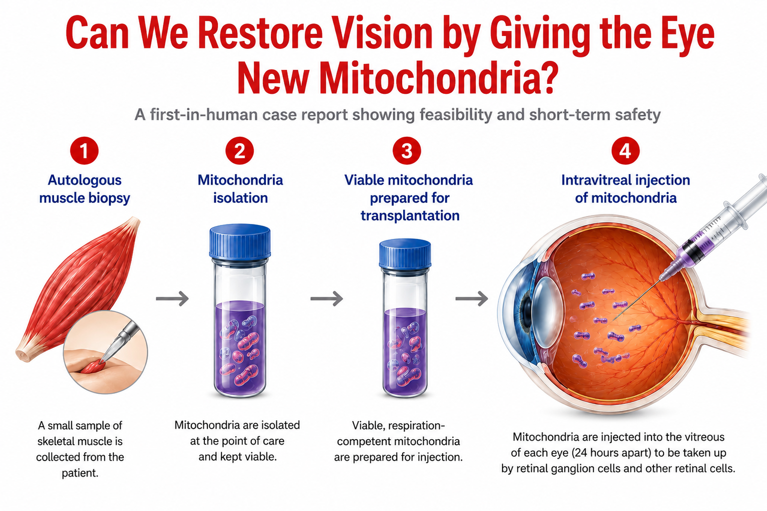
A first-in-human case explores whether viable autologous mitochondria can be delivered directly into the eye to rescue injured but still living neurons.
A new case report posted on Research Square describes what the authors report as the first administration of isolated mitochondria into the human eye.
David Putrino, Christopher Kellner, James McCully, Gareth Lema, Mansi Saxena, Joshua Bederson, Valerie Elmalem and Melanie Walker, from the Icahn School of Medicine at Mount Sinai, Boston Children’s Hospital and the University of Washington, report this first-in-human experience.
The study marks an important step in the emerging field of mitochondrial transplantation: delivering viable, respiration-competent mitochondria directly to tissues whose own mitochondrial function has been severely compromised.
From Mitochondrial Dysfunction to Mitochondrial Rescue
The patient was a 26-year-old woman who developed profound bilateral optic neuropathy following a severe intracerebral hemorrhage associated with prolonged cerebral hypoperfusion.
Approximately three months after the injury, visual function remained severely impaired and bilateral optic nerve atrophy was already evident.
Under an FDA-authorized individual-patient emergency expanded-access IND, the team isolated fresh mitochondria from the patient’s own skeletal muscle and injected them directly into the vitreous of each eye, 24 hours apart.
The biological rationale is compelling. Retinal ganglion cells have exceptionally high energy requirements and depend heavily on oxidative phosphorylation. Following ischemia-reperfusion injury, mitochondrial failure can contribute to ATP depletion, oxidative stress and ultimately neuronal death.
Mitochondrial transplantation does not aim to regenerate neurons that have already disappeared. Instead, it seeks to restore bioenergetic capacity in injured cells that remain viable.
That distinction may prove crucial. The success of mitochondrial transplantation could depend not only on the disease being treated, but also on when treatment is delivered and how much viable tissue remains to be rescued.
What Happened After the Injections?
The first important finding was feasibility and short-term safety.
Both injections were tolerated without serious adverse events. The investigators reported no intraocular inflammation, increased intraocular pressure, endophthalmitis, retinal detachment or detectable systemic inflammatory response.
There was also an intriguing functional signal.
During the 71 days immediately before treatment, none of 45 automated pupillometry readings across the two eyes reached the predefined threshold for a normal pupillary light reaction.
Following mitochondrial administration, normal-range responses appeared within days in both eyes. Overall, 13 of 52 post-treatment readings reached the threshold.
Organized occipital responses were also detected during post-treatment steady-state visual evoked potential assessments.
However, an important limitation must be emphasized: the response was transient and measurable visual acuity did not improve. The findings therefore suggest a physiological response, not restoration of vision.
Could Mitochondria Themselves Become Therapeutics?
This is perhaps the most important question raised by the report.
Mitochondrial transplantation has already begun moving from experimental models toward human investigation in cardiac and cerebral ischemia. This case extends the concept to another highly energy-dependent neuronal tissue: the retina and optic nerve.
Instead of using a drug to modify one mitochondrial pathway, organelle therapy introduces functioning biological machinery directly into metabolically compromised tissue.
This could represent a conceptual shift in mitochondrial medicine: from targeting mitochondria to considering mitochondria themselves as therapeutic agents.
WMS Scientific Board Perspective
Prof. Volkmar Weissig, Midwestern University, USA, sees the report as an important step for the field:
“The key question now is whether mitochondrial delivery can move from transient functional rescue toward a reproducible and durable therapeutic effect.”
For Prof. Marvin Edeas, Université Paris Cité, France, the implications could ultimately extend much further:
“What makes this report particularly interesting is that it may open a much wider therapeutic door. In many diseases, the cell is not immediately lost; it remains alive but energetically compromised because its mitochondria are failing. Stem-cell therapies aim mainly to replace damaged or lost cells. Mitochondrial transplantation raises another possibility: rescuing cells before they cross the point of no return.”
If this principle can eventually be demonstrated in controlled studies, the potential field of investigation could extend beyond the eye to stroke and cerebral ischemia, myocardial ischemia, retinal and optic nerve disorders, skeletal muscle injury and potentially some neurodegenerative conditions in which mitochondrial dysfunction contributes to cellular failure.
But major barriers remain.
“The next challenge is to determine which diseases are truly driven by mitochondrial dysfunction, which cells remain salvageable, and when the therapeutic window closes. We also need to understand how mitochondria should be delivered, at what dose, and whether repeated administration can produce a durable clinical benefit,” note Marvin Edeas and Volkmar Weissig.
These questions will determine whether mitochondrial transplantation can move beyond individual experimental applications and become a broader platform for organelle therapy.
A First Step, Not Yet Proof
The study cannot establish efficacy. It involves only one patient, has no control group, the functional changes were temporary, and visual acuity remained essentially unchanged.
Nevertheless, it provides an important first demonstration that fresh autologous mitochondria can be prepared at the point of care and administered intravitreally without the ocular or systemic toxicity anticipated as major potential concerns in this case.
The next challenge is no longer simply whether mitochondria can be delivered.
It is to determine where, when and in whom mitochondrial transplantation can produce meaningful and durable clinical benefit.
Reference: Putrino D, Kellner C, McCully J, et al. First intravitreal mitochondrial transplantation for bilateral vision loss. Research Square. Posted August 10, 2026. DOI: 10.21203/rs.3.rs-10622019/v1.
Scientific notice: This is a single-patient preprint case report that has not yet undergone peer review. Its findings should therefore be considered preliminary.
For more Information:
Read the full news here
Read the news on LinkedIn